What is Diagnostic Laparoscopy?
Diagnostic laparoscopy is a procedure that helps surgeon to see inside your abdomen, a hollow tube (port) is placed through your abdominal wall, and the laparoscopy is inserted into the port. The image of the inside of your abdomen is then seen on the monitor. In most cases, this procedure (operation) will be able to diagnose or help discover what is abdominal problem.
Why diagnostic laparoscopy performed?
Diagnostic laparoscopy is done for various reasons some of are listed here:
Abdominal pain
Laparoscopy has a role in the diagnosis of both acute and chronic abdominal pain. There are many causes of abdominal pain. Some of these causes include appendicitis, adhesions or intra-abdominal scar tissue, pelvic infections, endometriosis, abdominal bleeding and, less frequently, cancer. It is used in patients with irritable bowel disease to exclude other causes of abdominal pain. Surgeons can often diagnose the cause of the abdominal pain and, during the same procedure, correct the problem.
Abdominal mass
A patient may have a lump (mass or tumor), which can be felt by the doctor, the patient, or seen on an X-ray. Most masses require a definitive diagnosis before appropriate therapy or treatment can be recommended. Laparoscopy is one of the techniques available to your physician to look directly at the mass and obtain tissue to discover the diagnosis.
"Second look" procedure or cancer staging
Your doctor may need information regarding the status of a previously treated disease, such as cancer. This may occur after treatment with some forms of chemotherapy or before more chemotherapy is started. Also, information may be provided by diagnostic laparoscopy before planning a formal exploration of the abdomen, chemotherapy or radiation therapy.
Ascites
The presence of fluid in the abdominal cavity is called ascites. Sometimes the cause of this fluid accumulation cannot be found without looking into the abdominal cavity, which can often be accomplished with laparoscopy.
Liver disease
Non-invasive imaging techniques such as ultrasound, CT scan (computed tomography) and MRI (magnetic resonance imaging) may discover a mass inside or on the surface of the liver. If non-invasive imaging cannot give your physician enough information, a liver biopsy may be needed to establish the diagnosis. Diagnostic laparoscopy is one of the safest and most accurate ways to obtain tissue for diagnosis. In other words, it is an accurate way to collect a biopsy to sample the liver or mass without actually opening the abdomen.
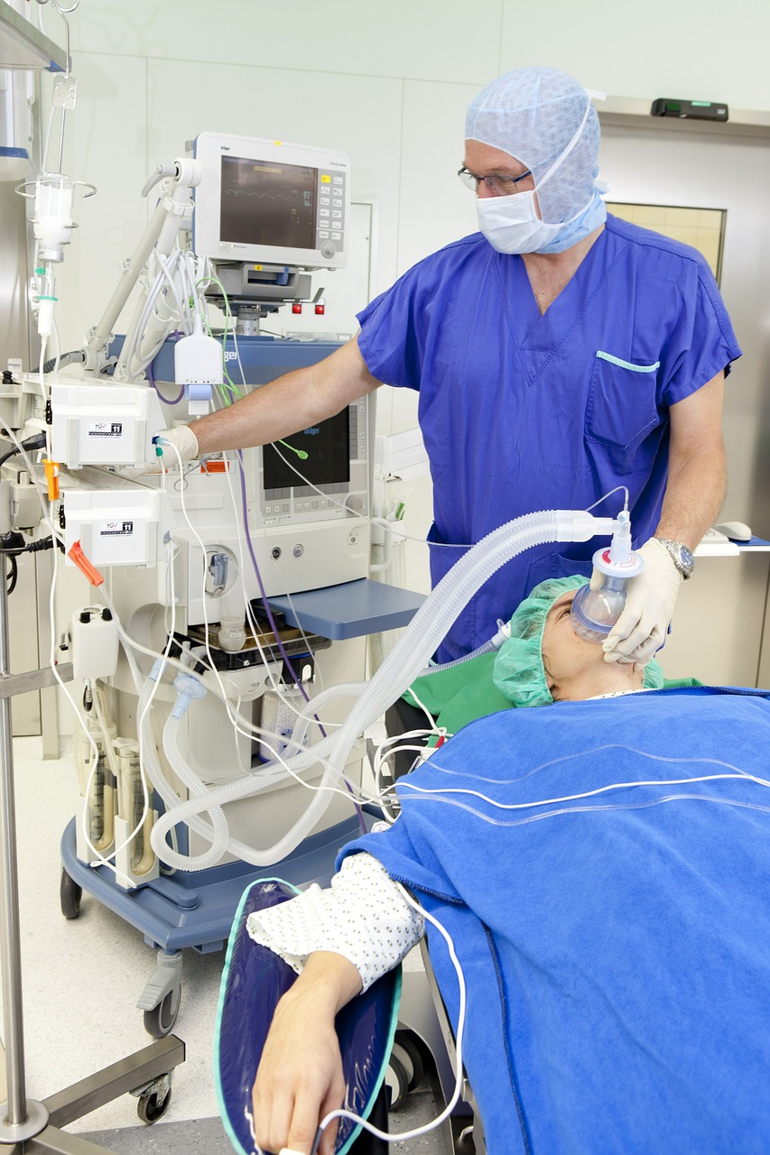
How is laparoscopy performed?
Laparoscopy is mainly done as an outpatient procedure. This implies you'll have the option to return home the same day as after surgery. It might be performed in a hospital or an outpatient surgical center.
You'll likely be given general anesthesia for laparoscopy surgery. This implies you'll rest through the operation and won't feel any pain. To accomplish general anesthesia, an intravenous (IV) line is embedded in one of your veins. Through the IV, your anesthesiologist can give special drugs and well as give hydration liquids.
In few cases, local anesthesia is used. A local anesthetic numbs the affected area, so despite the fact that you'll be conscious during the surgery, you won't feel any pain.
During laparoscopy surgery, the surgeon makes an incision beneath your belly button, and afterward embeds a tiny tube called a cannula. The cannula is used to inflate the abdomen with carbon dioxide gas. This gas permits surgeon to see your abdominal organs all the more clearly.
When abdomen is inflated, the surgeon embeds the laparoscope through the incision. The camera connected to the laparoscope shows the pictures on a screen, permitting your organs to be seen continuously.
The number and size of incision relies on what explicit disease your surgeon is endeavoring to affirm or preclude. Mainly, you get from one to four cuts that are each somewhere in the range of 1 and 2 centimeters long. These cuts permit other instruments to be embedded. For instance, your surgeon may need to use another surgical apparatus to do a biopsy. During a biopsy, they take a small sample of tissue from an organ to be assessed.
After the surgery is done, the instruments are expelled. Incisions are then shut with sutures or surgical tape. Bandages might be put over the incisions.
Hysteroscopy
Diagnostic Hysteroscopy is a procedure which allows doctor to look inside your uterus to diagnose and treat abnormal bleeding causes. It's study of the womb's inside using a fine telescope. A small telescope is inserted into the womb's cavity via the vagina and cervix. This is done usually under general anesthesia. The surgeon then investigates the lining of the womb carefully; the images from a camera attached to the telescope are projected onto a TV screen so you can watch the images if you wish. You'll probably need a biopsy of the womb lining. This is done by having a tiny sampler inserted at the end of the operation.
Diagnostic Laparoscopy V/s Hysteroscopy
Diagnostic laparoscopy may be recommended to look outside the uterus, Falopian tubes, ovaries, and internal pelvic area. Seeing inside the uterine cavity is used with diagnostic hysteroscopy.
 Help! just A Call Away