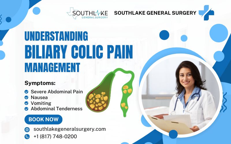
Biliary colic pain is a sharp and intense abdominal pain that occurs due to a blockage in the bile duct. This blockage prevents the flow of bile from the gallbladder to the small intestine, leading to discomfort and severe pain.
Gallstones or other obstructions can cause this condition, making it important to seek proper management and treatment for relief.
Understanding the causes, symptoms, and management options for biliary colic is crucial for individuals who experience this condition. This blog will provide an overview of biliary colic, including its causes, risk factors, and treatment options.
Key Highlights
- Biliary colic is a type of abdominal pain that occurs when a gallstone blocks the bile duct.
- The gall bladder plays a role in digestion by storing and releasing bile, which helps digest fats.
- Gallstones are the primary cause of biliary colic, but other factors such as obesity and liver disease can also contribute.
- Symptoms of biliary colic include severe abdominal pain, nausea, and vomiting.
- approaches for biliary colic include physical examination and imaging tests like ultrasound and CT scans.
- Treatment options for biliary colic include non-surgical management strategies and surgical interventions.
- Diet and lifestyle modifications, as well as pain management medications and therapies, can help manage biliary colic pain.
- It’s important to seek medical attention if you experience symptoms of biliary colic or if you have complications like acute cholecystitis or inflammation of the pancreas.
- FAQs: How can I prevent biliary colic attacks? Can diet alone manage biliary colic pain? What are the long-term implications of biliary colic?
Understanding Biliary Colic: An Overview
Biliary colic is a type of abdominal pain that occurs when a gallstone blocks the bile duct. The gallbladder, which stores and releases bile, plays a key role in digestion. When a gallstone obstructs the bile duct, the gallbladder contracts, causing pain and discomfort.
Other contributing factors to biliary colic include obesity, liver disease, and hormonal imbalances. Symptoms of biliary colic can be severe and may include abdominal pain, nausea, and vomiting. Prompt medical attention is necessary to diagnose and manage biliary colic effectively.
What is Biliary Colic?
Biliary colic is a type of abdominal pain that occurs when a gallstone blocks the bile duct. The bile duct is responsible for carrying bile from the gallbladder to the small intestine, where it helps with the digestion of fats. When a gallstone or other obstruction blocks the flow of bile, it can lead to intense pain and discomfort.
The pain associated with biliary colic typically occurs in the middle to the right upper abdomen and can radiate to the shoulder. It may be described as sharp, crampy, or a constant dull ache.
Biliary colic often occurs after eating a heavy meal, and the pain may last for 30 minutes to an hour or longer. It subsides when the gallstone passes into the intestine and unblocks the duct.
Biliary colic should not be confused with cholecystitis, which is inflammation of the gallbladder. Cholecystitis is a more severe condition that requires immediate medical attention.
If you experience symptoms of biliary colic, it is important to seek medical evaluation to determine the underlying cause and appropriate treatment.
The Role of the Gallbladder in Digestion
The gallbladder has a vital function in the process of digestion as it stores and releases bile. Bile is a hepatic secretion that helps in the breakdown and assimilation of dietary fats. After a meal, the gallbladder contracts, releasing bile into the small intestine through the bile ducts.
When we consume a meal that contains fats, the gallbladder contracts to release bile into the small intestine. Bile helps break down fats into smaller molecules, allowing for easier digestion and absorption. This process ensures that the body can utilize the nutrients from the fats we consume.
Without the gallbladder, bile would flow directly from the liver into the small intestine, bypassing the storage and concentration functions of the gallbladder. Although the gallbladder is not essential for digestion, its removal can affect the storage and release of bile, leading to changes in the digestion and absorption of fats. This can sometimes result in individuals experiencing symptoms such as diarrhea or fatty stools after gallbladder removal.
Understanding the role of the gallbladder in digestion is important in managing conditions such as biliary colic and making informed decisions about treatment options.
Causes and Risk Factors for Biliary Colic
Biliary colic is primarily caused by gallstones, which are hard masses that form in the gallbladder or bile ducts. Gallstones can consist of cholesterol, bilirubin, or a mixture of both.
Other factors that can contribute to the development of biliary colic include strictures or narrowing of the bile ducts and biliary dyskinesia, which is the impaired movement of bile through the biliary tract.
Certain risk factors increase the likelihood of developing gallstones and biliary colic, including being female, being over the age of 40, having a family history of gallstones, being overweight or obese, and having certain medical conditions such as liver disease or diabetes.
Gallstones: The Primary Culprit
Gallstones are the primary cause of biliary colic. These small, hard stones can form in the gallbladder or bile ducts. They are typically composed of cholesterol or bilirubin, a pigment produced by the breakdown of red blood cells.
Gallstones can vary in size and number, ranging from tiny sand-like particles to larger stones that can block the flow of bile. The formation of gallstones occurs when there is an imbalance in the bile components, such as an excess of cholesterol or bilirubin.
Certain factors increase the risk of developing gallstones, including obesity, rapid weight loss, a high-fat diet, and certain medical conditions such as liver disease and diabetes.
Women are also more prone to gallstone formation due to hormonal changes, particularly during pregnancy or with the use of estrogen-based contraceptives.
Other Contributing Factors
In addition to gallstones, several other factors can contribute to the development of biliary colic.
- Obesity is a significant risk factor, as excess body weight can lead to increased cholesterol levels in the bile, promoting the formation of gallstones.
- Liver disease, such as cirrhosis or hepatitis, can impair the normal flow of bile and increase the risk of gallstone formation.
- Hormonal factors, particularly estrogen, can also play a role in gallstone development.
- Women who are pregnant, taking hormone replacement therapy, or using oral contraceptives that contain estrogen are more likely to develop gallstones.
- Estrogen increases cholesterol levels in bile and reduces gallbladder contractions, which can contribute to the formation of gallstones and biliary colic.
Understanding these contributing factors can help individuals take preventative measures and manage their risk of developing biliary colic.
Identifying Symptoms of Biliary Colic
Identifying biliary colic symptoms is crucial for prompt diagnosis and treatment. Symptoms include severe abdominal pain in the upper right abdomen or beneath the ribs, which may be sharp, crampy, or a dull ache. Pain can radiate to the shoulder or back and often follows heavy meals, lasting hours.
Other symptoms include nausea, vomiting, and abdominal tenderness. Seek immediate medical attention if pain persists or is accompanied by fever, jaundice, or severe symptoms, as it could signal complications like cholecystitis or pancreatitis.
Common Signs and Symptoms
The most common symptom of biliary colic is severe abdominal pain, typically located in the upper right quadrant of the abdomen. The pain is often described as a severe, crampy, or squeezing sensation and can radiate to the shoulder or back.
The pain may come on suddenly and be intense, causing the individual to seek medical attention. This intense pain is often referred to as a “gallbladder attack.”
Nausea and vomiting are also common symptoms of biliary colic, as the intense pain can cause these digestive disturbances. It is important to note that the pain associated with biliary colic is different from other types of abdominal pain, such as indigestion or gas, as it is typically more severe and persistent.
If you encounter these symptoms, it is crucial to promptly seek medical attention for accurate diagnosis and appropriate treatment.
When to Seek Medical Attention
While biliary colic is usually not a medical emergency, there are instances where immediate medical attention should be sought. If the abdominal pain is severe, persistent, or accompanied by fever, chills, or jaundice, it may indicate complications such as acute cholecystitis or inflammation of the pancreas.
Acute cholecystitis is a condition where the gallbladder becomes inflamed and infected, requiring urgent medical intervention. Inflammation of the pancreas, known as pancreatitis, can also occur as a result of biliary colic and may lead to serious complications.
Additionally, if the pain is so severe that it interferes with daily activities or if you have a history of gallbladder disease, it is important to seek medical attention for proper diagnosis and treatment.
Prompt medical care can help prevent complications and ensure appropriate management of biliary colic.
Diagnostic Approaches

Diagnosing biliary colic requires a thorough medical history, physical examination, and various diagnostic tests. The process starts with obtaining a detailed medical history to understand symptoms and risk factors, followed by a physical examination to check for abdominal tenderness or swelling.
Imaging tests like ultrasound, CT scan, or MRI may be conducted to confirm the diagnosis by detecting gallstones, evaluating the gallbladder and bile ducts, and ruling out other potential causes of abdominal pain.
Sometimes, additional tests like endoscopic retrograde cholangiopancreatography (ERCP) are necessary for a more detailed view of the biliary tract.
Initial Assessment and Physical Examination
During the initial assessment, a healthcare provider will:
- Take a detailed medical history, including symptoms and risk factors for biliary colic
- Ask about the nature of the pain, its location, severity, and any associated symptoms like nausea or vomiting
- Conduct a physical examination, which may involve palpating the abdomen for tenderness or swelling
- Measure blood pressure and other vital signs to assess overall health
- The physical examination will focus on the upper abdomen where the gallbladder is located
- The healthcare provider will look for signs of gallbladder problems, such as tenderness or swelling, indicating inflammation or obstruction
This initial assessment and physical examination will guide further diagnostic testing and treatment decisions.
Imaging Tests Used in Diagnosis
Imaging tests play a crucial role in the diagnosis of biliary colic. The most common imaging test used is an ultrasound, which uses sound waves to create images of the gallbladder and bile ducts.
Ultrasound can help identify the presence of gallstones, assess the condition of the gallbladder, and detect any abnormalities in the bile ducts.
In some cases, additional imaging tests such as CT scan or MRI may be ordered to provide more detailed images of the biliary tract. These tests can help identify the location and extent of any abnormalities, such as gallstones or strictures.
In certain cases, endoscopic retrograde cholangiopancreatography (ERCP) may be performed. This procedure involves passing a flexible tube through the mouth and into the bile ducts to directly visualize the biliary tract and obtain images or tissue samples if needed.
Treatment Options for Biliary Colic
The treatment options for biliary colic depend on the underlying cause and the severity of symptoms. In most cases, surgical removal of the gallbladder, known as cholecystectomy, is the standard treatment for gallstones and biliary colic.
This procedure can be performed laparoscopically, using small incisions and a camera-guided instrument, or through open surgery in more complex cases. Cholecystectomy is a highly effective treatment, as the gallbladder is not essential for normal digestion.
Non-surgical management strategies may be used for individuals who are not suitable candidates for surgery or who have mild symptoms.
These strategies may include pain medication to manage symptoms, dietary modifications to reduce the risk of gallstone formation, and close monitoring of symptoms.
Non-Surgical Management Strategies
Non-surgical management strategies may be considered for individuals who are not suitable candidates for surgery or who have mild symptoms of biliary colic. These strategies aim to manage symptoms and reduce the risk of complications.
Pain medication, such as nonsteroidal anti-inflammatory drugs (NSAIDs), may be prescribed to alleviate pain during episodes of biliary colic.
Before taking any medication, it is crucial to adhere to the prescribed dosage and get guidance from a healthcare professional. Dietary modifications may also be recommended to reduce the risk of gallstone formation. This may involve avoiding fatty and high-cholesterol foods, eating a balanced diet rich in fruits, vegetables, and whole grains, and maintaining a healthy weight.
Regular follow-up with a healthcare provider is essential to monitor symptoms and receive appropriate medical care. In some cases, individuals may also be advised to undergo regular imaging tests to check for the presence of biliary sludge, which can increase the risk of gallstone formation.
Surgical Interventions

Surgical intervention is the most common and effective treatment for biliary colic caused by gallstones. The standard surgical procedure for gallstone removal is cholecystectomy, which involves the removal of the gallbladder.
This procedure can be performed using minimally invasive techniques, such as laparoscopic surgery, or through open cholecystectomy in more complex cases.
- Laparoscopic surgery involves making several small incisions in the abdomen and using a camera-guided instrument to remove the gallbladder. This approach offers several benefits, including shorter recovery time, less post-operative pain, and smaller incisions.
- Open cholecystectomy may be necessary if there are complications or if laparoscopic surgery is not feasible. It involves making a larger incision in the abdomen to remove the gallbladder.
The choice of surgical intervention depends on various factors, including the individual’s overall health, the severity of symptoms, and the presence of any complications.
Diet and Lifestyle Modifications for Management

Diet and lifestyle modifications can play a significant role in managing biliary colic and reducing the risk of gallstone formation. It is important to follow a healthy and balanced diet that is low in saturated fats and cholesterol. This involves avoiding or limiting the consumption of fatty and fried foods, processed foods, and foods high in cholesterol.
Instead, prioritize the inclusion of a greater variety of fruits, vegetables, whole grains, and lean meats in your dietary intake. Regular physical activity can also help maintain a healthy weight and promote overall digestive health.
Strive to engage in at least 30 minutes of exercise with a moderate level of intensity on most days of the week. It is also important to stay hydrated and limit the consumption of alcohol, as excessive alcohol intake can increase the risk of gallstone formation.
Foods to Avoid and Include
When managing biliary colic, it is important to be mindful of the foods you consume. Specific food items have the potential to elicit symptoms or actively contribute to the development of gallstones. Here are some tips for biliary colic pain management:
- Avoid or limit fatty foods, especially those high in saturated and trans fats, such as fried foods, fatty cuts of meat, full-fat dairy products, and processed snacks.
- Focus on consuming a balanced diet rich in fruits, vegetables, whole grains, lean proteins, and healthy fats from sources such as avocados and nuts.
- Stay hydrated by drinking plenty of water throughout the day.
By following a nutritious diet and making healthy choices, you can support your overall digestive health and manage biliary colic symptoms.
Importance of Physical Activity

Regular physical activity is essential for managing biliary colic and reducing the risk of gallstone formation. Physical activity plays a crucial role in maintaining a healthy weight, which is important as obesity is a significant risk factor for gallstone formation.
Engaging in regular exercise can help prevent weight gain and promote weight loss if necessary. It can also improve digestion and promote bile flow, reducing the risk of gallstone attacks and the formation of gallbladder sludge.
On most days of the week, try to exercise for at least 30 minutes at a moderate level. This can include such as brisk walking, cycling, swimming, or participating in a fitness class.
Remember to consult with a healthcare provider before starting any new exercise program, especially if you have any underlying health conditions.
Managing Pain: Medications and Therapies

Managing pain is an important aspect of biliary colic treatment. Medications can help alleviate pain during episodes of biliary colic and reduce discomfort.
Nonsteroidal anti-inflammatory drugs (NSAIDs) such as ibuprofen or naproxen be used to relieve pain and reduce inflammation. However, it is important to follow the recommended dosage and consult a healthcare provider before taking any medication.
In addition to medication, certain therapies such as heat therapy or relaxation techniques may help manage pain and promote relaxation.
- Applying a heating pad or hot water bottle to the affected area can provide temporary relief and promote muscle relaxation.
- Relaxation techniques such as deep breathing exercises or meditation can also help manage pain and reduce stress.
Over-the-Counter Pain Relief
Over-the-counter pain relievers can provide temporary relief from the abdominal pain associated with biliary colic. Nonsteroidal anti-inflammatory medicines (NSAIDs) like ibuprofen or naproxen can effectively soothe pain and decrease inflammation. These drugs function by suppressing the synthesis of specific molecules in the body that induce pain and inflammation.
It is important to follow the recommended dosage and consult a healthcare provider before taking any medication, especially if you have any underlying medical conditions or are taking other medications.
NSAIDs should be used for short-term relief of pain and should not be relied upon as a long-term solution. If pain persists or worsens, it is important to seek medical attention for proper diagnosis and treatment.
Prescription Medications
In some cases, prescription medications may be necessary to manage the symptoms of biliary colic. These medications are typically prescribed by a healthcare provider and are tailored to the individual’s specific needs.
For example, if inflammation of the gallbladder is present, medications such as antibiotics or anti-inflammatory drugs may be prescribed to reduce inflammation and alleviate pain.
Medications that help dissolve gallstones, such as ursodeoxycholic acid, may also be prescribed in certain cases. These medications work by modifying the composition of bile, making it less likely for gallstones to form.
Additionally, medications that promote the contraction of the gallbladder, such as cholecystokinin, may be prescribed to enhance the flow of bile and reduce the risk of gallstone formation.
It is important to follow the prescribed dosage and consult a healthcare provider for proper guidance and monitoring.
Outlook
The outlook for individuals with biliary colic is generally positive, especially with appropriate treatment and management. Surgical removal of the gallbladder, if necessary, is a highly effective treatment for biliary colic caused by gallstones. After gallbladder removal, most individuals can resume their normal activities without any significant impact on digestion or overall health.
Following a healthy lifestyle, including a balanced diet, regular exercise, and proper hydration, can help reduce the risk of gallstone formation and recurrence of biliary colic symptoms.
It is important to address any underlying risk factors, such as obesity or liver disease, to prevent future complications. Regular follow-up with a healthcare provider is essential to monitor symptoms and receive appropriate care for long-term management of biliary colic.
Make An Appointment

If you are experiencing symptoms of biliary colic or have been diagnosed with the condition, it is important to seek medical attention for proper evaluation and treatment.
Dr. Valeria Simone MD, a highly experienced general surgeon at Southlake General Surgery in Texas, USA, specializes in the treatment of gallbladder diseases and biliary colic. To schedule an appointment with Dr. Valeria Simone MD, please call +1 (817) 748-0200.
Dr. Simone and her team are committed to providing personalized and comprehensive care to help manage your biliary colic symptoms and improve your quality of life. Don’t delay seeking medical care for biliary colic. Contact Southlake General Surgery today to schedule an appointment.
Frequently Asked Questions
How can I prevent biliary colic attacks?
To prevent biliary colic attacks, it is important to maintain a healthy lifestyle. This includes following a balanced diet that is low in saturated and trans fats, maintaining a healthy weight, staying hydrated, and getting regular exercise. Avoiding or limiting the consumption of fatty and fried foods can also reduce the risk of gallstone formation. It is also important to manage any underlying risk factors, such as obesity or liver disease, as they can increase the likelihood of biliary colic.
Can diet alone manage biliary colic pain?
While diet plays a crucial role in managing biliary colic, it may not be sufficient to manage the pain alone. Dietary modifications, such as avoiding fatty foods and maintaining a balanced diet, can help reduce the risk of gallstone formation. However, for individuals with severe or persistent symptoms, additional medical interventions may be necessary, such as surgical removal of the gallbladder.
Can biliary colic pain lead to complications if left untreated?
If biliary colic pain is left untreated, it can lead to complications such as gallbladder inflammation, infections, or even blockage of the bile ducts. Seeking timely medical intervention and proper pain management strategies are essential to prevent such complications and ensure overall well-being.
What are the long-term implications of biliary colic?
The long-term implications of biliary colic depend on the underlying cause and the management of the condition. In most cases, surgical removal of the gallbladder is a highly effective treatment, and individuals can lead a normal and healthy life without their gallbladder. However, it is important to address any underlying risk factors and receive regular follow-up care to prevent complications and ensure overall well-being.
Medically Reviewed By: Dr. Valeria Simone MD
Board-certified General Surgeon at Southlake General Surgery, Texas, USA.
Follow us on Facebook and YouTube.
References:
- Pisano M, Allievi N, Gurusamy K, et al. 2020 World Society of Emergency Surgery updated guidelines for the diagnosis and treatment of acute calculus cholecystitis. World J Emerg Surg. 2020;15(1):61. Published 2020 Nov 5. https://doi:10.1186/s13017-020-00336-x
- Ansaloni L, Pisano M, Coccolini F, et al. 2016 WSES guidelines on acute calculous cholecystitis [published correction appears in World J Emerg Surg. 2016 Nov 4;11:52. doi: 10.1186/s13017-016-0088-z]. World J Emerg Surg. 2016;11:25. Published 2016 Jun 14. https://doi:10.1186/s13017-016-0082-5
- Kim E, Bashir R, Lahham S. Point-of-care Ultrasound Diagnosis of Cholecystitis vs. Adenomyomatosis. Clin Pract Cases Emerg Med. 2019;3(2):158-159. Published 2019 Mar 27. https://doi:10.5811/cpcem.2019.2.40798
- Dimou FM, Adhikari D, Mehta HB, Riall TS. Outcomes in Older Patients with Grade III Cholecystitis and Cholecystostomy Tube Placement: A Propensity Score Analysis. J Am Coll Surg. 2017;224(4):502-511.e1. doi:10.1016/j.jamcollsurg.2016.12.021
- Kim SY, Yoo KS. Efficacy of preoperative percutaneous cholecystostomy in the management of acute cholecystitis according to severity grades. Korean J Intern Med. 2018;33(3):497-505. doi:10.3904/kjim.2016.209
- “Acute Cholecystitis.” StatPearls – NCBI Bookshelf, 22 May 2023, www.ncbi.nlm.nih.gov/books/NBK459171.
- Fraquelli M, Casazza G, Conte D, Colli A. Non-steroid anti-inflammatory drugs for biliary colic. Cochrane Database Syst Rev. 2016;9(9):CD006390. Published 2016 Sep 9. doi:10.1002/14651858.CD006390.pub2
- Duncan CB, Riall TS. Evidence-based current surgical practice: calculous gallbladder disease. J Gastrointest Surg. 2012;16(11):2011-2025. doi:10.1007/s11605-012-2024-1
- Pak M, Lindseth G. Risk Factors for Cholelithiasis. Gastroenterol Nurs. 2016;39(4):297-309. doi:10.1097/SGA.0000000000000235
- Ansaloni L, Pisano M, Coccolini F, et al. 2016 WSES guidelines on acute calculous cholecystitis [published correction appears in World J Emerg Surg. 2016 Nov 4;11:52. doi: 10.1186/s13017-016-0088-z]. World J Emerg Surg. 2016;11:25. Published 2016 Jun 14. doi:10.1186/s13017-016-0082-5
- Shakespear, Jonathan S., et al. “CT Findings of Acute Cholecystitis and Its Complications.” American Journal of Roentgenology, vol. 194, no. 6, June 2010, pp. 1523–29. https://doi.org/10.2214/ajr.09.3640.
- “Gallstone Disease: Diagnosis and Management of Cholelithiasis, Cholecystitis and Choledocholithiasis.” PubMed, 1 Oct. 2014, pubmed.ncbi.nlm.nih.gov/25473723.
- Duncan, Casey B., and Taylor S. Riall. “Evidence-Based Current Surgical Practice: Calculous Gallbladder Disease.” Journal of Gastrointestinal Surgery, vol. 16, no. 11, Nov. 2012, pp. 2011–25. https://doi.org/10.1007/s11605-012-2024-1.