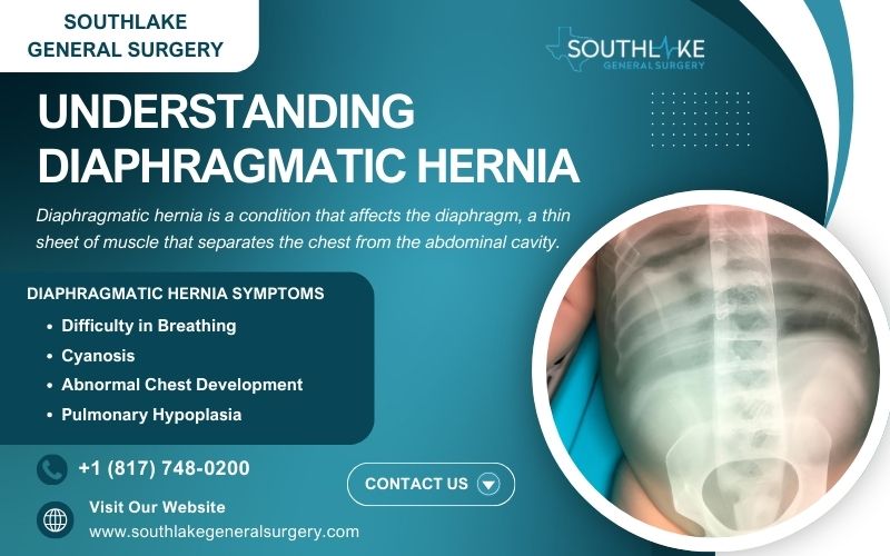
Diaphragmatic hernia is a condition that affects the diaphragm, a thin sheet of muscle that separates the chest from the abdominal cavity. In normal development, the diaphragm is fully formed by 10 weeks of gestation.
However, in cases of diaphragmatic hernia, there is an opening in the diaphragm, allowing abdominal organs such as the bowel, stomach, liver, and spleen to move upward into the chest cavity as the fetus develops. The medical term for this issue is congenital diaphragmatic hernia (CDH).
CDH is a birth defect that occurs in approximately 1 in every 3,000 live births. Although it often manifests on the left side, it is also possible to find it in the center or on the right.
CDH can have serious implications for the baby’s health, as the presence of abdominal organs in the chest limits the space available for the lungs to develop properly. This can lead to breathing complications after birth, which can be life-threatening.
One of the symptoms of congenital diaphragmatic hernia (CDH) is when the baby’s internal organs, such as the liver, intestines, or stomach, are in the baby’s chest instead of the lungs. Another indication is when the baby’s heart is pushed to one side by these additional organs.
The exact cause of CDH is not fully understood, but it is believed to be a combination of genetic and environmental factors. In some cases, CDH can be associated with genetic disorders or chromosomal abnormalities.
Yet, a hereditary component is not always present when CDH manifests. It is important to note that CDH is not caused by anything the mother did or did not do during pregnancy, but rather by a defect in the development of the esophagus.
Key Highlights
- Congenital diaphragmatic hernia (CDH) occurs when there is an opening in the diaphragm, allowing abdominal organs to move into the chest cavity.
- CD affects approximately 1 in 3,000 live births and most commonly occurs on the left side.
- The two types of diaphragmatic hernia are Bochdalek hernia and Morgagni hernia, with Bochdalek hernia being the most common.
- CDH can lead to breathing complications due to the limited space for lung development and can be life-threatening for newborns.
- Severity of CDH varies; parents should seek out specialist care promptly upon diagnosis.
Introduction to Diaphragmatic Hernia
Diaphragmatic hernia is a condition where abdominal organs move into the chest through an abnormal opening in the diaphragm. This occurrence can compress the developing lungs, leading to underdevelopment and associated complications.
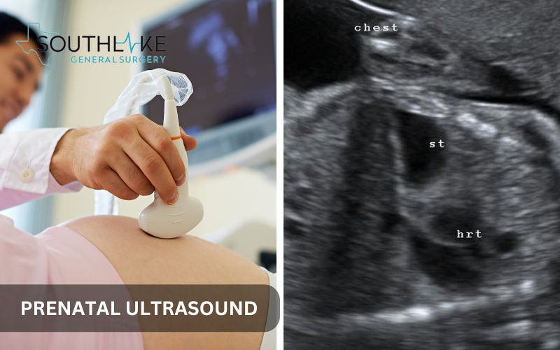
The most severe cases of congenital diaphragmatic hernia (CDH) can result in pulmonary hypoplasia, pulmonary hypertension, and persistent pulmonary hypertension in newborns. Although routine prenatal ultrasounds can sometimes detect the issue, the diagnosis may also occur after birth.
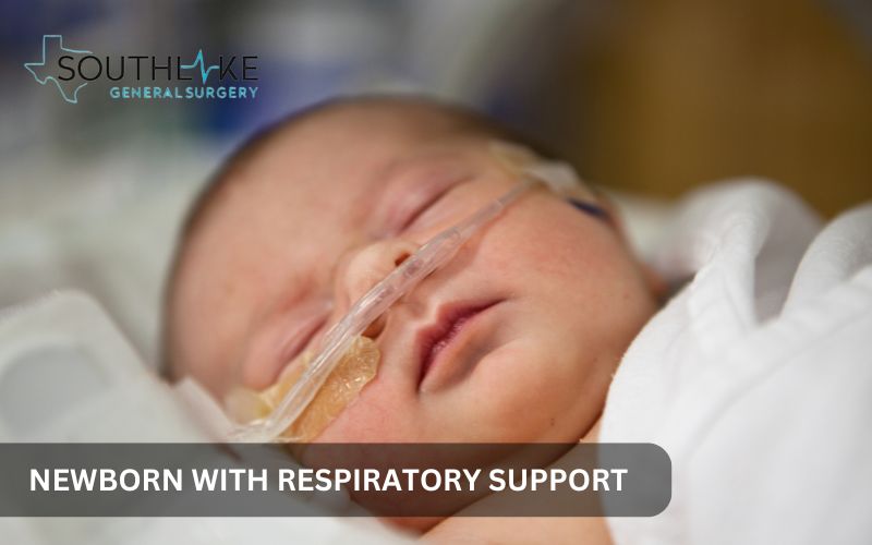
In some instances, treatment involves extracorporeal membrane oxygenation (ECMO) and mechanical ventilation to support respiratory function and manage associated difficulties.
Understanding the complexities of diaphragmatic hernias and the baby’s condition is crucial for timely intervention and better outcomes.
Defining Diaphragmatic Hernia
A diaphragmatic hernia occurs when there is an abnormal opening in the diaphragm, the muscle that separates the chest cavity from the abdominal cavity. This opening allows organs from the abdomen, such as the stomach, intestines, or liver, to move into the chest cavity.
This displacement can restrict lung development and lead to potentially life-threatening complications. Diaphragmatic hernias can either be congenital, present at birth, or acquired later in life due to trauma or surgery.
The most common type is a congenital diaphragmatic hernia (CDH), which affects lung growth and function in newborns. Understanding the nature of this condition is crucial in determining the appropriate treatment approach.
Defining congenital diaphragmatic hernia (CDH)
Congenital diaphragmatic hernia (CDH) is a birth issue. It causes an unusual hole in the diaphragm. This hole lets belly organs go into the chest, affecting lung growth. CDH can lead to small lungs and problems like high blood pressure in the lungs.
Finding it early through ultrasounds before birth or after birth is important for good care. Treatment might need ECMO and a machine to help with breathing. Knowing about CDH helps treat it on time and improves results for babies affected by it.
Different Types of Diaphragmatic Hernias
Diaphragmatic hernias can be either congenital (present at birth) or acquired (obtained later in life). Congenital diaphragmatic hernia (CDH) occurs when the lungs are not fully developed, resulting in the protrusion of abdominal organs into the chest cavity.
It is often diagnosed during routine prenatal ultrasounds. In the most severe cases of CDH, pulmonary hypoplasia and pulmonary hypertension can occur. Acquired diaphragmatic hernias are usually caused by trauma or surgery.
The treatment approach varies for each type, with CDH often requiring specialized care, including extracorporeal membrane oxygenation (ECMO) and mechanical ventilation.
Symptoms of Diaphragmatic Hernia
The symptoms of diaphragmatic hernia can vary depending on the severity of the condition. Some common symptoms include:
- Difficulty in breathing: Babies with diaphragmatic hernia may have trouble breathing immediately after birth. This is due to the limited space for lung development and the compression of the lungs by the abdominal organs in the chest cavity.
- Fast breathing: Rapid or shallow breathing can be a sign of respiratory distress in babies with this type of hernia.
- Cyanosis: Bluish discoloration of the skin, lips, or nails can indicate a lack of oxygen in the blood, which is a serious symptom that requires immediate medical attention.
- Abnormal chest development: In some cases, the chest may appear caved in or have an asymmetrical shape due to the displacement of abdominal organs.
If you notice any of these symptoms in your baby, it is important to seek medical attention immediately to ensure prompt diagnosis and treatment.
Recognizing Early Signs
Recognizing the early signs of diaphragmatic hernia can help in early diagnosis and management. Here are a few initial indicators to be aware of:
- Trouble breathing: Infants with diaphragmatic hernia may experience respiratory challenges shortly after delivery. This can manifest as rapid or labored breathing, flaring of the nostrils, or retractions (visible pulling in of the chest or abdomen with each breath).
- High blood pressure: Due to limited space for lung development and pulmonary hypoplasia, babies with diaphragmatic hernia may experience high blood pressure in the arteries of the lungs (pulmonary hypertension). This can further impair lung function and oxygenation.
- Pulmonary hypoplasia: Diaphragmatic hernia can lead to underdevelopment of the lungs, known as pulmonary hypoplasia. This can result in smaller-than-expected lungs with fewer air sacs (alveoli), limiting the baby’s ability to breathe properly.
If you notice any of these early signs, it is important to seek immediate medical attention. Early intervention and specialized care can significantly improve outcomes for babies with this type of hernia.
When to Seek Medical Attention
If you suspect that your baby may have a diaphragmatic hernia, it is important to seek medical attention as soon as possible. Early diagnosis and proper medical care are crucial for the well-being of the baby.
Contact your health care provider or seek emergency medical care if you notice any of the following signs or symptoms:
- Severe respiratory distress: Fast breathing, cyanosis (bluish discoloration of the skin), or difficulty breathing that worsens over time.
- Abnormal chest movement: Visible retractions (pulling in of the chest or abdomen with each breath) or flaring of the nostrils.
- Poor feeding or difficulty feeding: Babies with this medical condition hernia may have difficulty coordinating breathing and feeding, leading to poor weight gain.
- Severe CDH: In cases of severe diaphragmatic hernia, immediate medical intervention is necessary to stabilize the baby’s condition and provide specialized care.
Don’t hesitate to reach out to your healthcare provider if you have any concerns or questions regarding your baby’s health. They can guide you through the necessary steps for diagnosis and treatment.
Causes and Risk Factors

The causes of diaphragmatic hernia are not always clear, but there are some known risk factors that may increase the likelihood of developing the condition.
- Birth defect: Diaphragmatic hernia is considered a congenital birth defect, meaning it occurs during fetal development.
- Genetic factors: In some cases, this type of hernia may be associated with genetic disorders or chromosomal abnormalities.
- Environmental factors: Certain environmental factors, such as maternal exposure to certain medications or toxins, may increase the risk of diaphragmatic hernia.
It is important to note that this medical condition can occur spontaneously and without any identifiable cause. Further research is needed to fully understand the underlying causes of diaphragmatic hernia.
Understanding the Root Causes
Exactly what triggers diaphragmatic hernia is still a mystery. However, it is thought that this condition develops due to several causes.
- Birth defect: Diaphragmatic hernia is considered a congenital birth defect, meaning it occurs during fetal development. It is thought to result from a problem in the formation of the diaphragm, which leads to the opening through which abdominal organs can move into the chest cavity.
- Genetic factors: In some cases, this medical condition may be associated with genetic factors. Certain genetic disorders or chromosomal abnormalities can increase the risk of developing a diaphragmatic hernia.
- Environmental factors: While the exact environmental factors that contribute to this type of hernia are not yet known, there is evidence to suggest that maternal exposure to certain medications or toxins during pregnancy may increase the risk.
It is important to note that diaphragmatic hernia can occur spontaneously in some cases, without any identifiable genetic or environmental cause. Further research is needed to better understand the root causes of this condition.
Identifying High-Risk Groups
While diaphragmatic hernia can occur in any baby, there are certain risk factors and high-risk groups that should be aware of the increased likelihood of this condition.
- Family history: If there is a family history of diaphragmatic hernia or other congenital birth defects, the risk may be slightly higher.
- Genetic factors: Certain genetic disorders or chromosomal abnormalities can increase the risk of diaphragmatic hernia. If there is a known genetic condition in the family, the risk may be higher.
- History of affected pregnancies: If a prior pregnancy was impacted by a diaphragmatic hernia, there is a slightly higher probability of it happening again.
- Number of live births: Studies have shown that the risk of diaphragmatic hernia may be slightly higher in pregnancies with multiple gestations (twins, triplets, etc.).
It is important to discuss any known risk factors or concerns with your healthcare provider. They can provide personalized guidance and support throughout your pregnancy and help ensure the best possible outcome for you and your baby.
Diagnosing Diaphragmatic Hernia
Diagnosing diaphragmatic hernia involves a combination of prenatal and postnatal tests and evaluations. Early diagnosis is crucial for planning appropriate medical care and interventions for the baby.
- Prenatal ultrasound: Diaphragmatic hernia is frequently identified during the standard 20-week prenatal ultrasound examination. This imaging test allows the health care provider to visualize the structure of the developing fetus and identify any abnormalities, including diaphragmatic hernia.
- Additional prenatal tests: In some cases, additional tests, such as fetal echocardiogram and fetal MRI, may be performed to evaluate the severity of the diaphragmatic hernia and assess the function of the heart and other organs.
- Postnatal evaluations: After birth, the health care provider will conduct a physical examination and order further tests to confirm the diagnosis of a diaphragmatic hernia. These may include chest X-rays, arterial blood gas tests, and ultrasound of the heart (echocardiogram).
Early diagnosis and proper evaluation are essential for managing diaphragmatic hernia and providing the necessary medical interventions for the baby’s well-being.
Diagnostic Tests and Procedures

Diagnosing diaphragmatic hernia involves a series of tests and procedures to evaluate the extent of the condition and plan appropriate medical care. Here are a few examples of common diagnostic tests and procedures:
- Prenatal ultrasound: A standard 20-week prenatal ultrasound screening often detects diaphragmatic hernias. This imaging test allows the health care provider to visualize the developing fetus and identify any abnormalities, including diaphragmatic hernia.
- Fetal echocardiogram: The purpose of this ultrasound is to assess the health and development of the baby’s heart. It helps to determine if there are any cardiac abnormalities associated with the diaphragmatic hernia.
- Fetal MRI: In some cases, fetal magnetic resonance imaging (MRI) may be performed to provide detailed images of the baby’s chest and abdomen, allowing for a more comprehensive evaluation of the diaphragmatic hernia and associated abnormalities.
These diagnostic tests and procedures are important for determining the severity of the diaphragmatic hernia and planning appropriate medical interventions for the baby’s care.
Interpreting Diagnostic Results for Diaphragmatic Hernia
Interpreting the diagnostic results of a diaphragmatic hernia is crucial for understanding the severity of the condition and providing appropriate medical care. Some key factors that are evaluated include:
- Presence of abnormalities: Diagnostic tests such as prenatal ultrasound, fetal echocardiogram, and MRI can reveal any associated abnormalities or complications, such as heart defects or underdeveloped lungs.
- Ultrasound results: Ultrasound measurements, including the lung volume or the observed to expected lung-to-head ratio (o/e LHR), are used to evaluate pulmonary hypoplasia and diaphragmatic hernia.
- Prognosis: Given the diagnostic results, a healthcare provider can offer a prognosis for the baby. This includes discussing the potential challenges and long-term outcomes associated with this type of hernia.
Interpreting the diagnostic results requires expertise and specialized knowledge. It is important to consult with a healthcare provider who has experience in managing diaphragmatic hernia to ensure the best possible care for the baby.
Treatment Options for Diaphragmatic Hernia

The complexity of the illness and the unique requirements of the infant dictate the course of treatment for diaphragmatic hernia. The objective of treatment is to stabilize the infant’s condition, provide assistance to lung function, and, if required, perform repair on the diaphragmatic hernia.
- Non-Surgical Management: In some cases, non-surgical interventions may be used to support the baby’s breathing and nutritional needs. This can include mechanical ventilation, oxygen therapy, and the use of a feeding tube.
- Surgical Interventions and Innovations: For severe cases of this type of medical condition, surgical repair of the diaphragm may be necessary. Advanced surgical techniques, such as extracorporeal membrane oxygenation (ECMO) and minimally invasive thoracoscopic repair, may be used to improve outcomes.
The treatment plan will be tailored to the individual needs of the baby, and a multidisciplinary team of specialists will be involved in providing comprehensive care and support.
Non-Surgical Management
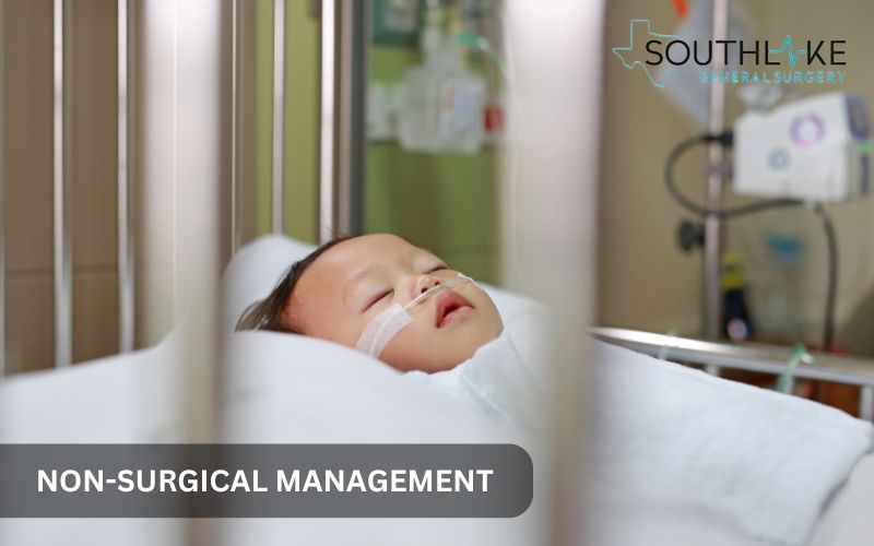
Non-surgical management plays a crucial role in the care of babies with diaphragmatic hernia, especially in the initial stabilization phase. Some common non-surgical interventions include:
- Mechanical ventilation: Babies with diaphragmatic hernia often require support from a mechanical ventilator to assist with breathing. This process facilitates the transportation of oxygen and elimination of carbon dioxide from the lungs.
- Oxygen therapy: Supplemental oxygen may be provided to maintain adequate oxygen levels in the blood and support the baby’s respiratory function.
- Feeding tube: Babies who have a this type of hernia may have trouble feeding since they can’t control their breathing as they eat. A feeding tube may be used to ensure adequate nutrition while minimizing the risk of aspiration.
Non-surgical management is aimed at optimizing lung function, maintaining adequate oxygenation, and supporting the baby’s nutritional needs. Regular monitoring and adjustments are made based on the baby’s condition and response to treatment.
Surgical Interventions and Innovations

When dealing with a serious diaphragmatic hernia, surgical procedures are essential. Significant advancements in surgical techniques and innovations have greatly enhanced outcomes for infants diagnosed with this condition. Thoroughly observing the following is essential after surgery:
- Extracorporeal membrane oxygenation (ECMO): In the most severe cases of this type of hernia, ECMO may be used as a temporary support system for the baby’s heart and lungs. It facilitates the removal of carbon dioxide from the blood and enhances the oxygenation process, aiding in the lungs’ recovery and repair.
- Minimally invasive surgery: The diaphragmatic hernia can be fixed using minimally invasive procedures like thoracoscopic repair. This method utilizes small incisions and specialized instruments, along with a camera, to carry out the surgical procedure.
These surgical interventions and innovations are aimed at repairing the diaphragmatic hernia, optimizing lung function, and improving outcomes for babies with severe diaphragmatic hernia. The approach used will vary based on the unique requirements of the infant and the proficiency of the surgical team.
Post-Surgery Care and Recovery
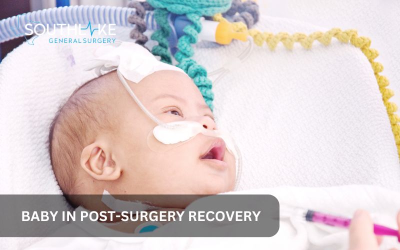
Post-surgery care and recovery are important aspects of the management of diaphragmatic hernia. Following surgical intervention, the baby will require close monitoring and support to ensure a smooth recovery. Thoroughly observing the following is essential after surgery:
- Hospital stay: Babies undergoing surgery for diaphragmatic hernia will typically require a hospital stay for further monitoring and management. Hospital stays can range from a few days to a few weeks, depending on factors including the baby’s individual needs and the extent of the operation.
- Follow-up appointments: Regular follow-up appointments will be scheduled to monitor the baby’s progress and address any concerns or complications that may arise. This may include imaging tests, developmental assessments, and consultations with specialists.
Post-surgery care and recovery are individualized based on the baby’s condition and the specific surgical interventions performed. The multidisciplinary care team will provide guidance and support throughout the recovery process.
What to Expect After Surgery
After surgery for a diaphragmatic hernia, there are several things to expect during the recovery process. Some key aspects include:
- Hospital stay: The infant will need to stay in the hospital for a while after surgery so they can receive extra care and observation. The length of the hospital stay will vary depending on the individual needs of the baby and the extent of the surgery.
- Recovery process: The recovery process will involve close monitoring of the baby’s vital signs, respiratory function, and overall well-being. Managing pain and caring for wounds are also crucial aspects of the healing process.
- Follow-up appointments: The baby’s progress and any problems or worries can be addressed during the regularly scheduled follow-up visits. These appointments may include imaging tests, developmental assessments, and consultations with specialists.
The recovery process will be individualized based on the baby’s condition and the specific surgical interventions performed. The care team will provide guidance and support throughout the recovery journey.
Long-Term Care Strategies
Long-term care strategies are important for babies who have undergone surgery for diaphragmatic hernia. Important components of long-term care comprise:
- Chronic lung disease: Babies with diaphragmatic hernia may be at risk of developing chronic lung disease, which may require ongoing monitoring and management. This may involve regular pulmonary function tests, medications to support lung function, and lifestyle modifications.
- Gastroesophageal reflux: Gastroesophageal reflux is common in babies with this type of hernia. It is important to manage reflux symptoms to prevent complications such as aspiration and poor weight gain. This may involve medications, changes in feeding techniques, and positioning strategies.
- Developmental delays: Babies with diaphragmatic hernia may be at risk of developmental delays, especially if they require ECMO support or experience other complications. Early intervention services, such as physical therapy, occupational therapy, and speech therapy, may be recommended to support the baby’s development.
Long-term care strategies will be individualized based on the baby’s specific needs and challenges. Regular follow-up appointments and open communication with the care team will help ensure comprehensive and tailored care for the baby.
Living with Diaphragmatic Hernia
Living with a diaphragmatic hernia requires ongoing care and monitoring to address potential long-term health issues and support the baby’s overall well-being. Some key aspects of living with diaphragmatic hernia include:
- Lifestyle adjustments: Depending on the severity of the diaphragmatic hernia, certain lifestyle changes may be recommended. This can include avoiding exposure to smoke, maintaining a healthy diet, and ensuring a safe and supportive environment for the baby.
- Regular monitoring: Regular check-ups and monitoring with the healthcare provider are crucial for assessing the baby’s growth, lung function, and overall health. This includes monitoring for any potential complications or long-term effects of the condition.
- Communication and support: Open communication with the healthcare team and seeking support from family, friends, and support groups can help navigate the challenges of living with a this medical condition.
By actively managing the baby’s health, staying informed about the condition, and seeking appropriate support, it is possible to provide the best possible quality of life for babies with diaphragmatic hernia.
Outlook
The outlook for babies with diaphragmatic hernia can vary depending on the severity of the condition and the presence of any associated complications. Some key factors that can influence the outlook include:
- Survival rate: The overall survival rate for babies with this type of hernia has significantly improved over the years. Advances in medical interventions and specialized care have contributed to higher survival rates, particularly in specialized centers with expertise in managing diaphragmatic hernia.
- Quality of life: While the prognosis for survival has improved, babies with diaphragmatic hernia may still face long-term health challenges. These can include respiratory issues, developmental delays, gastroesophageal reflux, and potential complications related to surgery and other interventions.
- Long-term outcomes: Each individual’s long-term outcomes can vary depending on the severity of this type of hernia, the presence of associated complications, and the effectiveness of medical interventions. Regular monitoring, ongoing care, and early intervention services can help mitigate potential challenges and optimize long-term outcomes.
Talk to your doctor about your expectations for your baby’s future. They can provide personalized information and support based on the individual circumstances and needs of your baby.
Make An Appointment
If you would like to discuss diaphragmatic hernia or seek specialized care, you can make an appointment with Dr. Valeria Simone MD, at Southlake General Surgery in Texas, USA.
Dr. Simone is an experienced surgeon with expertise in the management of diaphragmatic hernia and can provide personalized care and guidance for you and your baby.
If you would like to schedule an appointment, kindly reach out to our dedicated team at +1 (817) 748-0200. They will gladly offer the required support.
Frequently Asked Questions
How successful is diaphragmatic hernia surgery?
The success rate of diaphragmatic hernia surgery depends on various factors, including the severity of the condition, associated complications, and the expertise of the surgical team. Advances in surgical techniques and specialized care have significantly improved outcomes, with higher survival rates in specialized centers in the United States. It is important to consult with a qualified surgeon to discuss the specific success rate and potential outcomes for your baby.
Can diaphragmatic hernia reoccur after surgery?
While rare, diaphragmatic hernia can reoccur after surgical repair. The risk of recurrence depends on various factors, such as the size of the hernia, the presence of associated complications, and the effectiveness of the initial surgical repair. Regular follow-up appointments and long-term monitoring are essential to detect any potential recurrence and address it promptly if it occurs.
How can I prevent complications associated with diaphragmatic hernia?
Preventing complications associated with diaphragmatic hernia involves early diagnosis, specialized medical care, and lifestyle adjustments. Timely prenatal diagnosis and appropriate medical management can help optimize outcomes. Following the recommended treatment plan, regular follow-up appointments, and making necessary lifestyle adjustments can help reduce the risk of complications and support the baby’s health and well-being.
How common is a diaphragmatic hernia in adults versus children?
Diaphragmatic hernia is more common in infants, with congenital diaphragmatic hernia occurring in about 1 in every 2,500 to 5,000 live births. In contrast, diaphragmatic hernia in adults is rare, often resulting from trauma or surgery. Regular check-ups and early diagnosis are crucial for both age groups.
Medically Reviewed By: Dr. Valeria Simone MD
Board-certified General Surgeon at Southlake General Surgery, Texas, USA.
Follow us on Facebook and YouTube.
References:
- Bielinska, Malgorzata, et al. “Molecular genetics of congenital diaphragmatic defects.” Annals of Medicine, vol. 39, no. 4, Jan. 2007, pp. 261–74. https://doi.org/10.1080/07853890701326883.
- Schumacher, Lana, and Sebastien Gilbert. “Congenital Diaphragmatic Hernia in the Adult.” Deleted Journal, vol. 19, no. 4, Nov. 2009, pp. 469–72. https://doi.org/10.1016/j.thorsurg.2009.08.004.
- Gaxiola, Alejandra, et al. “Congenital diaphragmatic hernia: an overview of the etiology and current management.” Acta Paediatrica, vol. 98, no. 4, Mar. 2009, pp. 621–27. https://doi.org/10.1111/j.1651-2227.2008.01212.x.
- Holder, A. M., et al. “Genetic Factors in Congenital Diaphragmatic Hernia.” The American Journal of Human Genetics, vol. 80, no. 5, May 2007, pp. 825–45. https://doi.org/10.1086/513442.
- Scott, Daryl A. “Genetics of congenital diaphragmatic hernia.” Seminars in Pediatric Surgery, vol. 16, no. 2, May 2007, pp. 88–93. https://doi.org/10.1053/j.sempedsurg.2007.01.003.
- Kantarci, Sibel, and Patricia K. Donahoe. “Congenital diaphragmatic hernia (CDH) etiology as revealed by pathway genetics.” American Journal of Medical Genetics Part C Seminars in Medical Genetics, vol. 145C, no. 2, Apr. 2007, pp. 217–26. https://doi.org/10.1002/ajmg.c.30132.
- Wat, M. J., et al. “Genomic alterations that contribute to the development of isolated and non-isolated congenital diaphragmatic hernia.” Journal of Medical Genetics, vol. 48, no. 5, Apr. 2011, pp. 299–307. https://doi.org/10.1136/jmg.2011.089680.
- Keijzer, Richard, and Prem Puri. “Congenital diaphragmatic hernia.” Seminars in Pediatric Surgery, vol. 19, no. 3, Aug. 2010, pp. 180–85. https://doi.org/10.1053/j.sempedsurg.2010.03.001.
- Klaassens, M., et al. “The etiology of congenital diaphragmatic hernia: Still largely unknown?” European Journal of Medical Genetics, vol. 52, no. 5, Sept. 2009, pp. 281–86. https://doi.org/10.1016/j.ejmg.2009.05.005.
- Yu, Lan, et al. “Variants in GATA4 are a rare cause of familial and sporadic congenital diaphragmatic hernia.” Human Genetics, vol. 132, no. 3, Nov. 2012, pp. 285–92. https://doi.org/10.1007/s00439-012-1249-0.